
The spinal cord itself extends through the entire cervical spine. 36 MRI can provide information about the soft tissues, including the neural tissue (spinal cord and nerve roots. Sometimes this measurement is included in a report. MRI is the modality of choice for early diagnosis of cervical involvement, because it has high sensitivity in detecting inflammatory changes in the jointssynovial changes and pannus formationeven before instability develops. Some report even larger, up to 16 millimeters. The level of this lecture is appropriate for medical students, junior residents, and trainees in other specialties who have an interest in neuroradiology or may see patients with spine diseases. Medical Animations by Certified Medical Illustrations.For additional information, please visit our website at or call toll-free 1-888-999. The normal cervical spine canal should be between 10-14 millimeters.
#Mri of cervical spine how to
As we walk through, we’ll take a look at how to use each one. The images can be stored on a computer or printed on film. In general, it is made of a lower and an upper part.

The optimal approach is to use select sequences to evaluate each part of the study in the following order:Įach sequence in the study has strengths at looking at one or more of these things. A cervical MRI (magnetic resonance imaging) scan uses energy from strong magnets to create pictures of the part of the spine that runs through the neck area (cervical spine). A cervical spine (CS) coil is usually more raised so that it provides better support for the patients neck. Before ordering Magnetic Resonance Imaging (MRI scan) for a patient with back pain or neck pain, the physician will usually consider the following general.

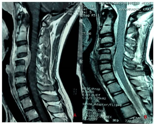
Magnetic resonance, several sagittal images searching for disk disease. This video will walk you through a step-by-step approach to evaluating an MRI of the cervical spine. MR image of cervical spine, all segments - diagnostic radiology - showing disk herniation (C5-C6 level). Most of these cases will be done without contrast, as most of the information is there on a non-contrast exam. This can include the spine, the space around the spinal cord, and vertebrae in your neck. An MRI can give your doctor information about the spine in your neck (the cervical spine). Magnetic resonance imaging (MRI) of the cervical spine is a very commonly encountered test which can be performed for a variety of indications, including degenerative disease, trauma, demyelinating disease, and metastatic disease. MRI (magnetic resonance imaging) is a test that uses a magnetic field and pulses of radio wave energy to make pictures of the organs and structures inside the body. Of a dog with a plasmocytoma on Octoin Heidelberg, Germany. x-ray right foot broken metatarsus fixed with screw - mri cervical spine stock illustrations. Noncontrast MRI cervical spine search pattern magnetic resonance images of cervical spine sagittal t2-weighted images (mri cervical spine) - mri cervical spine stock pictures, royalty-free photos & images.


 0 kommentar(er)
